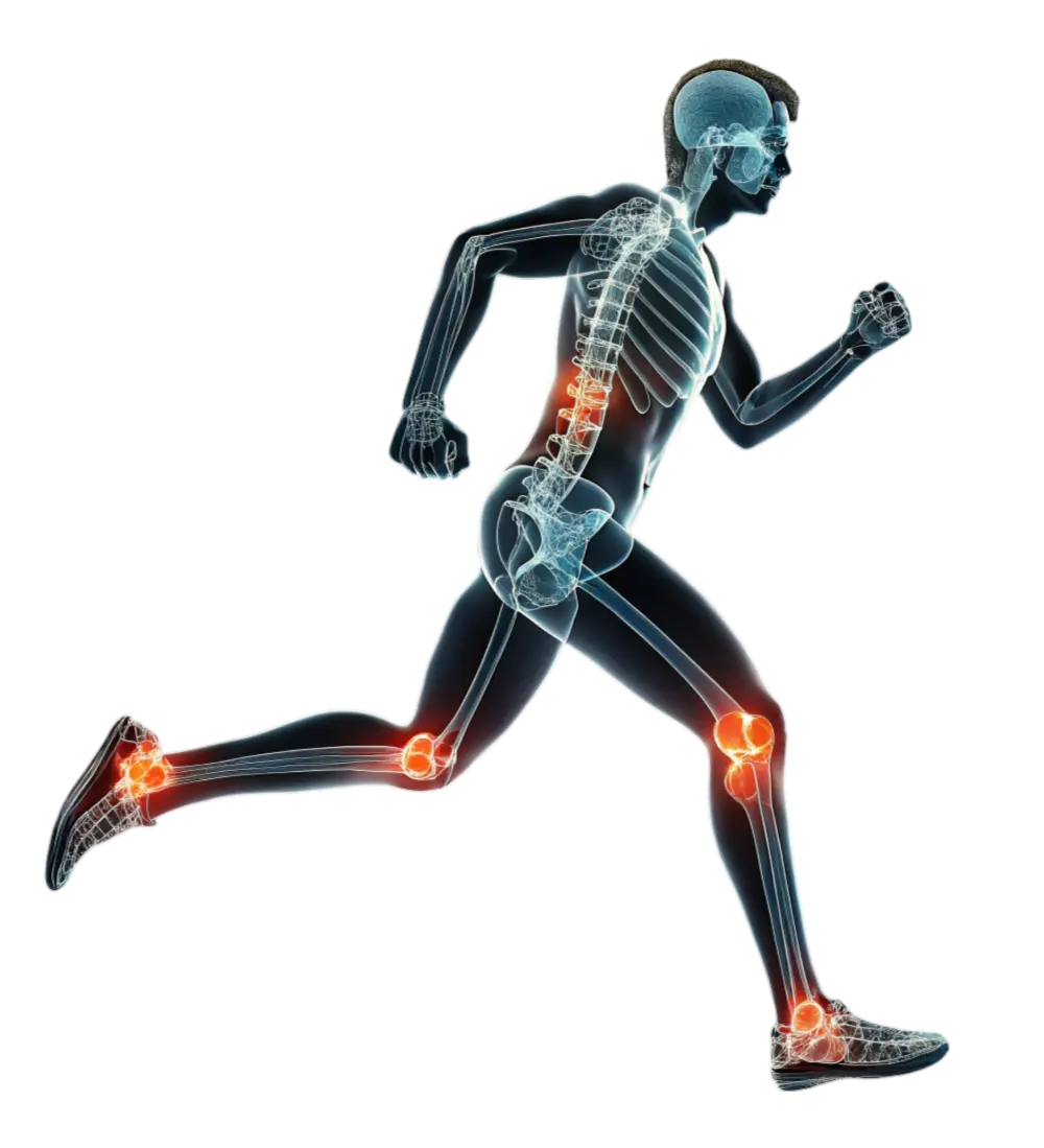
MRI stands for magnetic resonance imaging. MRI is used by physicians to look inside the body without using X-rays or radiation. It uses safe magnetic fields, radiowaves, and computers to produce detailed and precise images of specific areas of the body, including the muscles, bones, and joints. MRI provides an accurate diagnosis of many musculoskeletal abnormalities and, in some cases, can reduce the need for biopsies, exploratory surgery, and other diagnostic procedures.
MRI is a painless and noninvasive diagnostic procedure with no known side effects or after effects. During the procedure, you won’t feel anything; you’ll simply hear a faint knocking sound during the actual imaging process.
During the procedure, you lie on a table inside the magnetic field, which aligns magnet-like protons in the body. When radio frequency waves are beamed through the magnetic field and absorbed by the protons, they then emit a signal, which is received and measured by a receiver coil. The computer then constructs an image of the body using these measurements. A specially trained, certified radiologist interprets the test results.
The procedure is painless and typically takes 25 to 30 minutes, depending on the type of information your doctor has requested. All you need to do is remain as still as possible during the exam. For your convenience, an intercom system is available should you need anything.
Nothing special is required prior to your MRI procedure. Simply follow your normal daily routine, including taking medication and meals. You may also want to bring your favorite CD or tape to play during the exam.
Because of the magnetic field, you should avoid wearing or carrying:
Bone x-ray uses a very small dose of ionizing radiation to produce pictures of any bone in the body. It is commonly used to diagnose fractured bones or joint dislocation. Bone x-rays are the fastest and easiest way for your doctor to view and assess bone fractures, injuries and joint abnormalities.
This exam requires little to no special preparation. Tell your doctor and the technologist if there is any possibility you are pregnant. Leave jewelry at home and wear loose, comfortable clothing. You may be asked to wear a gown.
Southern Bone & Joint Specialists now has digital x-ray capabilities. Instead of X-ray film, digital radiography uses a digital image capture device. This Diagnostic Imaging gives advantages of immediate image preview and availability; elimination of costly film processing steps; a wider dynamic range, which makes it more forgiving for over- and under-exposure; as well as the ability to apply special image processing techniques that enhance overall display quality of the image.